Introduction.
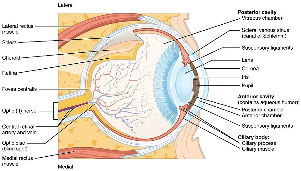
The human eyes are located in two small parts of the skull called orbits or eye sockets. The eyelids clean dirt from the eyes and protect them from dehydration. They produce tears in their eyes, which contain substances that fight bacterial infections. The eyelids prevent particles from entering the eyes. The structure is divided into three layers:
Layers:
- Outer layer ( sclera and cornea)
- Middle layer ( choroid)
- Inner layer ( retina)
1. Outer layer.
The outer layer of the human eye consiste of the two layers: Sclera and Cornea.
Sclera.
- The sclera gives the white color of the eye.
- It consiste of the connective tissue.
- They protect the inner component of the eye.
- They maintain the shape of the eye.
Corena.
- In the front, the Transparent layer formed by the sclera is called the Cornea.
- The cornea allows light to enter the eye.
- The cornea also bends the rays of light so that they come to a focus.
- Light focuses on the retina.
2. Middle layer.
The outer layer of the human eye consiste of the Choroid.
- Blood vessels are present. They give the dark color of the inner eye.
- The dark colour prevents disruptive reflections within the eye.
- Behind the cornea, the choroid bends to form a muscular ring, called the Iris.
- In the center of the iris is a round red pupil.
- After being refracted through the cornea, light passes through the pupil.
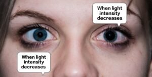
- The muscles of the iris adjust the size of the pupil.
- In bright light, the circular muscles of the iris contract and the pupil narrows.
- In dim light, the radial muscles of the iris contract and the pupil dilates.
- Behind the iris is a Convex lens. that focuses light onto the retina.
- The lens is attached to the Ciliary Muscles of the eye by a circular Suspensory Ligament.
- To see objects at a greater distance, the ciliary muscles relax and the lens becomes less convex.
- The contraction of the ciliary muscles causes the lens to become narrower and rounder.
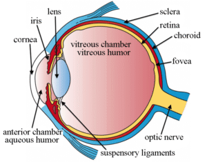
3. Inner layer.
The Inner layer of the human eye consiste of the Retina. It contains light-sensitive cells, namely Rods and Cones, and their associated neurons. Rods detect dim light (night vision). Cones detect bright light and color vision. The retina has two points: the Fovea and the Optic Disc.
Fovea.
- The fovea is a dip in the retina directly opposite the lens.
- The fovea has a very high number of cone cells.
- This area is responsible for color recognition and the sharpness of vision.
Optic Disc.
- The optic disc is the point on the retina where the optic nerve enters the retina.
- Rods and cones are not found at this point, so it is also called the blind spot.
The iris divides the eyeball into two chambers. The anterior chamber is in front of the iris, between the cornea and the iris, while the posterior chamber is between the iris and the retina. The anterior chamber contains a clear fluid called the Aqueous humor, while the posterior chamber contains a jelly-like fluid called the Vitreous humor. This helps maintain the shape of the eye and also keeps the delicate lens still. When light enters the eye from an object is refracted as it passes through the cornea, iris, lens, and vitreous humor. The lens also focuses this light on the retina, and as a result, the retina is imaged in the same way.
Light from objects enters the eye and is refracted when it passes through the cornea, aqueous humour, lens, and vitreous humour. The lens also focuses this light onto the retina, resulting in an image on the retina. Rods and cones produce nerve impulses in the optic nerve. These impulses are carried to the brain, which makes the sensation of vision.
A pigment is found inside the rods called Rhodopsin. When light falls on the rods, the nerve fibers break down to produce a pulse. In the absence of light, the broken products of the rods break down and form the rods. Our body makes the rods from Vitamins A, and this is the reason why we cannot see well at night due to a lack of vitamins. This disease is called Night Blindness.
Cones also contain a protein called Iodopsin. There are three major types of cones, and each type produces a specific type of iodine. Each type of cone has a color: blue, green, and red. If any one of the types of cones does not work properly, it becomes difficult to recognize that color. A person is also unable to distinguish between different colors. This disease is called Color Blindness and is a genetic disease.
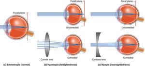
Disorders of the Eye:
Changes in the shape of the eyeball affect the function of the eye.
Myopia (Short-sightedness).
This defect occurs due to the curvature of the eyeball. Such people cannot see distant objects clearly. The focus of distant objects is formed in front of the retina. This defect can be corrected by using a concave lens.
Hypermetropia (Long sight).
This defect occurs due to the shortening of the length of the eyeball. Such people cannot see close objects clearly. The image of distant objects is formed behind the retina. This defect can be corrected by using a convex lens.