
Introduction.
Spermiogenesis is the final stage of spermatogenesis (the process of sperm cell formation) in which immature spermatids transform into mature, functional spermatozoa (sperm cells) without undergoing further cell division.
- Spermiogenesis is the final stage of spermatogenesis.
- It is the morphological differentiation of haploid round spermatids (non-motile, immature cells) into highly specialized and motile spermatozoa (sperm cells).
- Unlike earlier stages, no cell division occurs in spermiogenesis — only structural and functional changes.
Stages of Spermiogenesis.
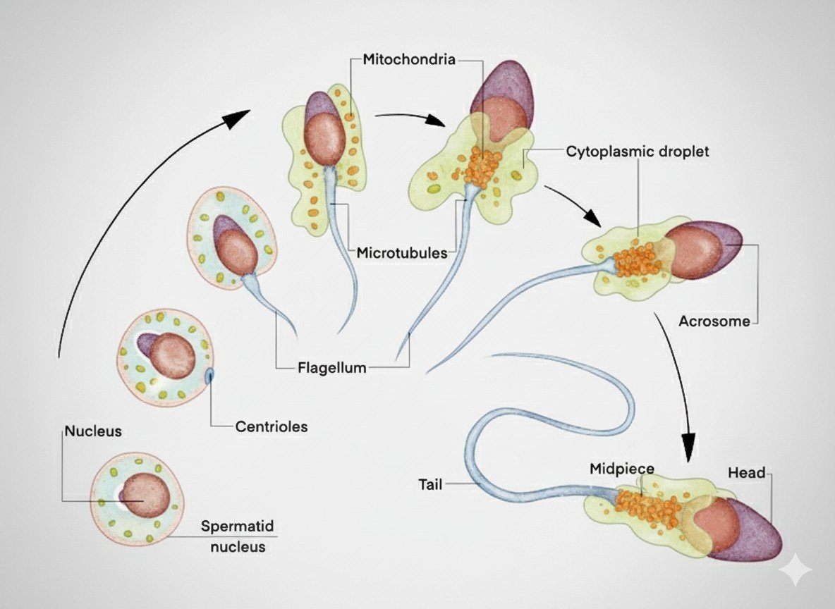
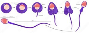
Spermiogenesis is the last stage of spermatogenesis in which spermatids develop into the mature spermatozoa. They complete into the four stages. The four stages are the Golgi phase, the cup phase, the Acrosome phase, and the maturation phase. Many different changes occur during these phases.
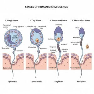
1. Golgi Phase
This is the first stage of spermiogenesis, where spermatids develop into sperm.
- The Golgi apparatus of the spermatid produces small proacrosomal vesicles.
- These vesicles fuse to form a large acrosomal vesicle, which attaches to the anterior part of the nucleus.
- Centrioles migrate to the posterior pole:
- Proximal centriole → connects sperm head to tail.
- Distal centriole → initiates growth of the axoneme, the core of the flagellum (tail).
- Result: Beginning of acrosome formation and tail initiation.
2. Cap Phase
The Cup phase is the second stage of spermiogenesis, in which the developing sperm is a more specialized structure.
- The acrosomal vesicle flattens and spreads over the anterior nuclear surface, forming the acrosomal cap.
- The acrosome contains hydrolytic enzymes like:
- Acrosin
- Hyaluronidase
- Neuraminidase
- These enzymes will help the sperm penetrate the egg’s protective layers during fertilization.
- The nucleus starts to condense and elongate.
3. Acrosome Phase
The acrosome is the third stage of spermiogenesis in which the spermatid changes its structure for several times and undergoes a significant change, and it looks like mature sperm.
Nuclear changes:
- The major changes occur in the nucleus and prepare for fertilization.
- The nucleus becomes highly condensed, elongated, and streamlined for better motility.
- The condensation makes the nucleus smaller.
- The size is reduced
Tail development:
- The flagellum elongates behind the nucleus.
- Axoneme structure develops (9 + 2 microtubule arrangement).
Mitochondria:
- Gather around the proximal part of the flagellum to form the mitochondrial sheath (middle piece).
- Provide ATP for sperm motility.
- The cytoplasm starts reducing, pushed toward the posterior.
4. Maturation Phase
- Excess cytoplasm forms residual bodies, which are phagocytosed by Sertoli cells.
- Final shaping of sperm occurs.
- Mature spermatozoa are released into the lumen of seminiferous tubules (a process called spermiation).
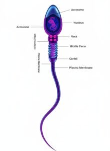
Final Structure of a Mature Spermatozoon.
Head
- Contains a condensed nucleus with haploid chromosomes.
- The acrosome covers the anterior head → has enzymes for oocyte penetration.
- The sperm head is not only a genetic material carrier but also a unique structure that is specific and necessary for reproduction and fertilization to occur
Neck
- The neck of a sperm is a short, connecting region that joins the head to the tail (flagellum). While small, the neck is extremely important for the structure and function of the sperm cell.
- Connects the head and middle piece.
- Contains centrioles that organize the tail microtubules.
- The neck is also important for movement because it assists in transferring energy generated in the middle piece of the sperm to the tail for motility.
Middle Piece
- The middle piece of the sperm is an important part of the sperm situated between its neck and tail (principal piece). Its main function is to provide the energy necessary to move.
- Contains mitochondria arranged helically around the axoneme.
- Supplies energy (ATP) for flagellar movement.
- The middle piece also has dense fibers to provide structural support to help the sperm maintain its shape and flexibility while moving.
Tail (Principal & End Piece)
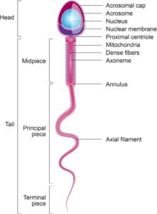
- Long flagellum with axoneme (9+2 microtubules).
- Provides motility, essential for reaching the oocyte.
- At the end of the tail is the end piece, which tapers off for directional guidance of the sperm. The next stage of movement behind the tail is in a coordinated fashion, powered by energy from the mitochondria, and aids the sperm’s use for optimal function through the female reproductive tract, which is then essential for fertilization.
Key Points.
- Spermiogenesis ≠ Spermatogenesis (it’s just the last phase).
- Takes place in the seminiferous tubules, inside the Sertoli cell support.
- Time required: ~ 24 days (in humans).
- Hormonal control:
- FSH → stimulates Sertoli cells.
- Testosterone (from Leydig cells) → essential for differentiation.


