
Introduction.
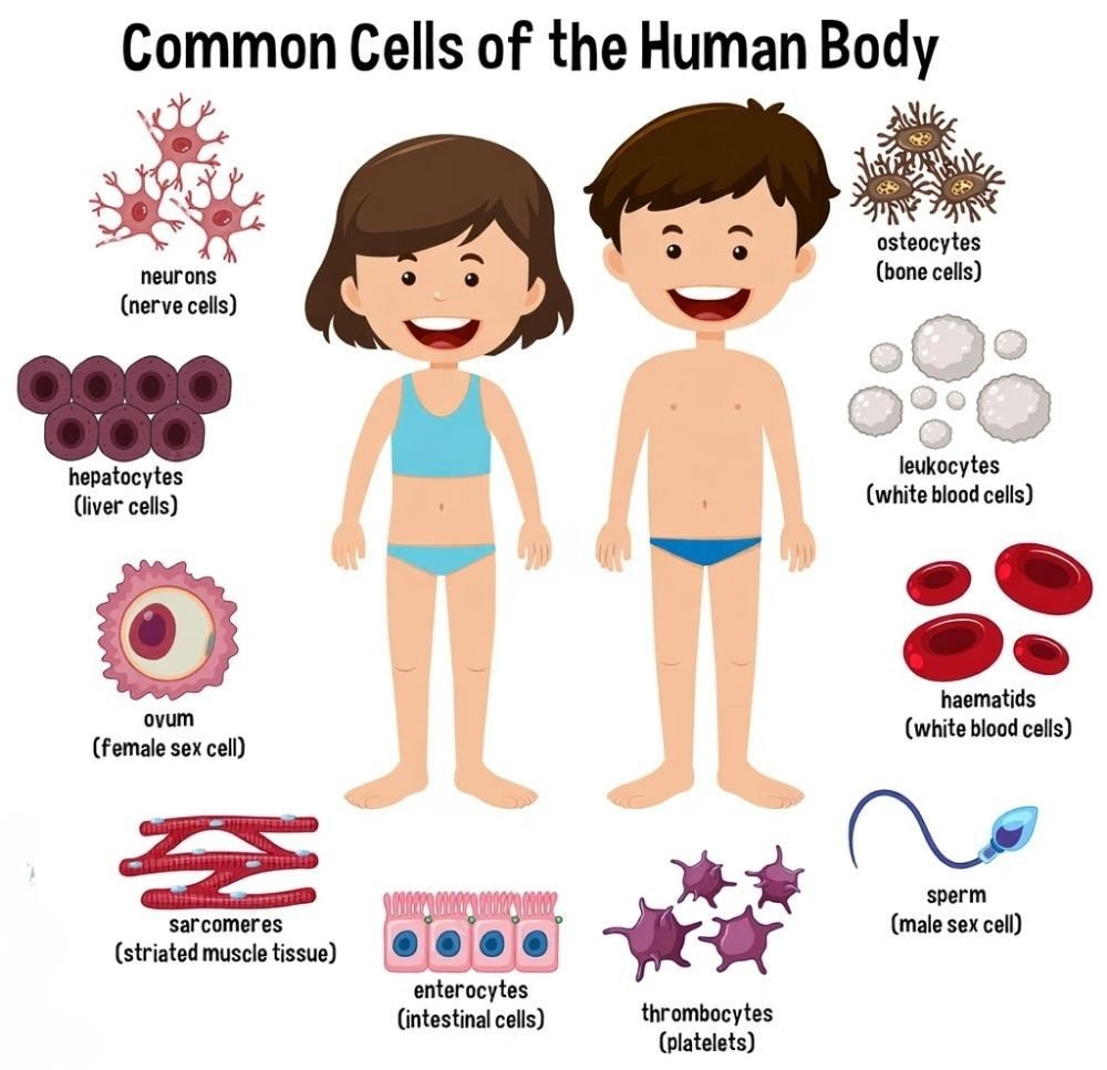
The human cell is the cell structure and functional unit of the human body that creates a foundation of life. Each human cell is a type of eukaryotic cell, meaning that there is a true nucleus that holds genetic material (DNA) and many membranes surrounded by organelles that have specialized functions. Human cells have a great deal of specialization and diversity. There are nerve cells that send signals, muscle cells that provide movement, blood cells that transport oxygen and fight infections and reproductive cells that are responsible for heredity. Despite different, characteristics employed they hold common functions of providing structure, producing energy, regulating genetic information, and allowing growth and repair. Trillions of these cells work together to sustain the health of the body and to ensure survival. Here are some different types of human cells.
Types:
- Red Blood Cells
- Cardiac Cells
- Sperm Cells
- Egg Cells
- Kidney Cells
- Smooth Muscle Cells
- Skeletal Muscle Cells
- Osteocyte Cells
- Neuron Cells
- Skin Cells
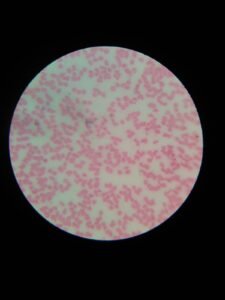
1. Red Blood Cells.
Red Blood Cells (RBCs), which are also known as erythrocytes, are the most numerous type of blood cell in the human body. They are specialized cells designed to deliver oxygen from the lungs to body tissues and bring carbon dioxide back to the lungs to be exhaled.
Structure of RBCs:
- Shape: Biconcave disc shape (providing larger surface area for gas exchange).
- Size: Almost 7–8 µm in diameter.
- Nucleus: Mature RBCs do not have a nucleus (which provides more space for fluid hemoglobin).
- Organelles: They have a less mitochondria (to avoid using the oxygen being transported).
- Color: RBSs have red color
- Lifespan: The lifespan is almost 120 days (4 months).
Functions of RBCs:
- RBCs transport oxygen in the whole body tissue by binding with the hemoglobin protein.
- They carry the carbon dioxide and release it in the lungs.
- Hemoglobin helps maintain the blood pH.
- RBCs’ shape provides flexibility to pass through small capillaries.
Formation:
- They form in the bone marrow by a process called erythropoiesis.
- The hormone erythropoietin is released from the kidneys, which stimulates production.
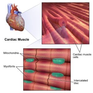
2. Cardiac Cells.
Cardiac cells are the specialized muscle cells that make up the heart. They form the cardiac muscle tissue (myocardium) and are responsible for the continuous pumping of blood throughout the body. Unlike skeletal muscles, cardiac cells contract involuntarily and rhythmically without conscious control.
Structure:
- Short, branched, and striated fibers.
- Usually, there is one centrally located nucleus.
- Intercalated discs connect cells, containing:
- Gap junctions (electrical signal transmission).
- Desmosomes (strong adhesion).
- Rich in mitochondria for a constant energy supply.
Function:
- Pump blood by contracting rhythmically.
- Generate and conduct impulses (pacemaker cells).
- Work continuously without fatigue.
- Coordinate contraction through intercalated discs.
Formation:
- Originate from mesodermal stem cells during development.
- After birth, they have very limited regeneration.
- Damage (e.g., heart attack) often leads to scar tissue instead of new cardiac cells.
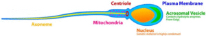
3. Sperm Cells
The sperm cell is the male gamete in sexual reproduction and it is responsible for transmitting the father’s genetic material to the female egg (ovum) during fertilization. It is also one of the smallest but most specialized cells within the human body
Structure of Sperm Cell:
- A sperm cell has a highly specialized structure adapted to its role in fertilization. A sperm cell has 3 main parts:
- Head Contains the nucleus with haploid DNA (23 chromosomes) and the acrosome, which has enzymes to penetrate the egg.
- The midpiece is packed with mitochondria that supply energy for movement.
- Tail (Flagellum) A long, whip-like structure that enables motility, helping the sperm swim toward the egg.
Functions of Sperm Cell:
- Delivers the father’s genetic material (DNA) to the egg.
- The Tail allows movement through the female reproductive tract.
- Enzymes help penetrate the protective layers of the egg.
- The sperm carries either an X or a Y chromosome, which determines the baby’s sex
Formation of Sperm (Spermatogenesis)
- Occurs in the seminiferous tubules of the testes.
- Begins at puberty and lasts throughout life.
- Sperms take 64–72 days to complete development.
- After development, sperm are held in to gain maturity in the epididymis until ejaculation.
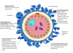
4. Egg Cells.
The egg cell, or ovum, is the female gamete in humans and is one of the largest cells in the body. It provides not only half of the genetic material (23 chromosomes) but also the cytoplasm, nutrients, and organelles necessary for the early stages of embryo development.
Structure of Egg Cell:
- In nucleus egg cell contains 23 chromosomes that are in the form of a haploid (n).
- The cytoplasm of egg cell is also called Ooplasm; they contains rich nutrients and organelles for egg development.
- Plasma membrane are present around the cytoplasm; they maintain the entry of sperm in the egg.
- Zona Pellucida – A thick glycoprotein layer that protects the egg and helps sperm bind.
- Corona Radiata – Outer layer of follicle cells surrounding the egg, providing nourishment.
Function of Egg Cell:
- Stores the mother’s genetic material.
- The egg cell provides energy rich material and cell metabolism for initial embryo development.
- In fertilization, a sperm cell enters the egg cell membrane, called the zona pellucida.
- Ensures species-specific fertilization and blocks entry of multiple sperm (polyspermy).
Formation of Egg Cells (Oogenesis)
- Begins before birth in females – oogonia develop into primary oocytes.
- Upon reaching puberty each monthly menstrual cycle, an oocyte becomes mature and is released during ovulation.
- The egg is viable for approximately 12 – 24 hours after ovulation.
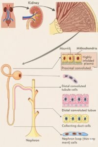
5. Kidney Cells.
Kidney cells are specialized to perform filtration, absorption, secretion, and regulation. Their structure varies depending on their location in the nephron.
structure of kidney cells:
- Kidney cells have different structures based on their function.
- Tubular epithelial cells are cuboidal/columnar with microvilli and many mitochondria for absorption and transport.
- Podocytes have foot-like processes (pedicels) forming filtration slits.
- Endothelial cells are fenestrated (porous) for filtration.
- Principal and intercalated cells in the collecting duct are cuboidal, with receptors or mitochondria for water, ion, and pH regulation.
function of kidney cells:
- Kidney cells are important to maintain the body’s internal balance.
- Filtration: Podocytes and glomerular endothelial cells filter blood, removing waste but keeping useful molecules.
- Reabsorption: Tubular epithelial cells reabsorb water, glucose, amino acids, and ions.
- Secretion: Certain cells secrete wastes, toxins, and hydrogen ions into urine.
- Regulation: Principal and intercalated cells regulate water, electrolytes, blood pressure, and pH balance under hormonal control (ADH, aldosterone).
Formation:
- Most kidney cells develop from the intermediate mesoderm during embryogenesis.
- Unlike skin or blood cells, kidney cells have very limited regeneration.
- Severe damage (e.g., chronic kidney disease) often leads to loss of function instead of repair.
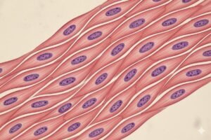
6. Smooth Muscle Cells.
Smooth muscle cells are spindle-shaped (elongated and tapered at both ends), uninucleate (having a single central nucleus), and non-striated (lack the visible bands seen in skeletal and cardiac muscle). They are found in the walls of hollow organs such as the stomach, intestines, blood vessels, urinary bladder, and uterus.
Structure:
- Spindle-shaped (elongated, tapered at ends).
- Single nucleus located in the center of the cell.
- Striations not visible (as compared to striated skeletal muscle and cardiac muscle);
- Has actin and myosin filaments arranged irregularly as the cause of slow and prolonged contraction
- Surrounded in connective tissue and connected with gap junctions for coordinated muscle cell activity.
Function:
- Found in the walls of hollow organs (intestines, stomach, blood vessels, bladder, uterus).
- Involuntary control (regulated by the autonomic nervous system & hormones).
- Functions include role in:
- controlling the diameter of the blood vessel (blood pressure)
- causing movement of food through the digestive tract (peristalsis)
- controlling airflow in the airways
- controlling the contraction of the uterus during childbirth
- controlling the emptying of the bladder.
Formation of Smooth Muscle Cells:
- Origin: Smooth muscle cells will originate mostly from mesoderm, (the middle germ layer) of the embryo.
- During development and mesodermal formation, Mesenchymal stem cells in the mesoderm will differentiate into myoblasts and progress and mature into smooth muscle cells.
- In adults, Smooth muscle cells can still divide and regenerate to some extent (unlike skeletal muscle). This regeneration is supported by resident stem cells (pericytes) located around blood vessels.
- Regulation: Their production and growth are influenced by growth factors (e.g., TGF-β, PDGF, FGF) and signals from surrounding tissues.
7. Skeletal Muscle Cells.
Skeletal muscle cells (muscle fibers) are long, cylindrical, striated, multinucleated cells. Skeletal muscle (muscle) refers to the skeletal muscle belonging to the skeletal system that is attached to bones and voluntary: moving facial, walking, and lifting.
Structure:
- Long, cylindrical, multinucleated cells (fibers).
- Show striations (alternating light and dark bands) due to organized actin and myosin filaments.
- Nuclei are located at the periphery of the cell.
- Each cell is packed with myofibrils, which contain sarcomeres (the contractile units).
Function:
- Factors in voluntary body movement (walking, lifting, posture).
- Create force through contraction in response to commands by the somatic nervous system.
- Also participate in heat generation (shivering)..
Formation:
- Formed from the mesodermal layer in embryonic life.
- Precursor cells termed myoblasts fuse to form long multinucleated fibers (syncytium).
- In adults most repair and limited regeneration is provided by satellite cells (muscle stem cells) located under the basal lamina.
8. Osteocyte Cells.
Osteocytes are mature bone cells found in lacunae that are primarily sensory and regulatory cells of bone health and develop when osteoblasts become trapped in the matrix of bone.
Structure:
- Osteocytes are the bone cells that arise from osteoblasts.
- They nest in lacunae (small voids in the bone matrix) and have long, star-shaped processes that communicate with other cells to provide a basis for nutrient and waste exchange.
- Osteocytes are moreover surrounded by a mineralized component of the matrix that prevents them from dividing.
Function:
- Osteocytes are responsible for the maintenance and monitoring of the integrity of the underlying bone/extracellular matrix (calcium and phosphate levels).
- They are sensitizers for detecting mechanical stress or pressure on the bone surface and signal nearby osteoblasts or osteoclasts for remodeling of the bone matrix.
- Help in the repair and long-term maintenance of bone tissue.
Formation:
- Osteocytes are produced when osteoblasts (bone-forming cells) become trapped in the bone matrix they secrete and differentiate into mature osteocytes.
- Osteocytes are mesodermal in origin (from mesenchymal stem cells).
9. Neuron Cells.
Neurons are a specific type of nerve cell that sends signals via electrical impulses and chemical signals to enable the brain and nervous system to perform their functions.
Structure:
- Neurons are the biological units of the nervous system.
- Having four main parts:
- the cell body (soma) storing the nucleus and organelles,
- the dendrites which are branch-like extensions that receive signals from other cells,
- the axon, which is a long fiber that sends impulsions away from the cell body, and the axon terminals joining up with other neurons and/or muscles or glands to send impulse signals to nearby neurons.
- Many axons are lined with a myelin sheath, coding the axon fiber and enabling faster communication.
Function:
- Neurons receive, process, and send electrical and chemical signals
- They form networks that transmit information and perform their functions for thought, memory, sensation, movement, or reflex.
- Communicate through synapses using neurotransmitters.
Formation:
- Neurons are derived from neuroectodermal cells of the embryonic ectoderm.
- During the formation of the nervous system neurons are produced from neural stem cells and neuroblasts.
- In non adult, only limited amounts of new neurons can be produced in certain regions of the brain (e.g., hippocampus) and even then at limited amounts.
10. Skin Cells.
The skin cells are keratinocytes, melanocytes, Langerhans cells, and Merkel cells, which all contribute to a common purpose of protecting the body, regulating body temperature, sensing the environment, and maintaining skin health.
Structure:
- The skin is the largest organ of the body and is made up of different cell types arranged in layers.
- Major types of skin cells include:
- Keratinocytes: Main cells of the epidermis; produce keratin for strength and protection.
- Melanocytes: Found in the basal layer; produce melanin (skin pigment).
- Langerhans cells: Immune cells that detect pathogens.
- Merkel cells: Sensory cells involved in touch sensation.
Function:
- Protection: Acts as a barrier against pathogens, UV radiation, and physical injury.
- Sensation: Detect touch, temperature, pain, and pressure.
- Regulation: Help maintain body temperature and prevent water loss.
- Pigmentation: Melanocytes provide skin color and protect from UV damage.
Formation:
- Skin cells are produced from stem cells in the basal layer of the epidermis.
- Old cells are pushed upward, filled with keratin, and eventually shed (takes about 28–40 days in humans).
- Melanocytes and Langerhans cells arise from different embryonic origins (neural crest and bone marrow, respectively).


