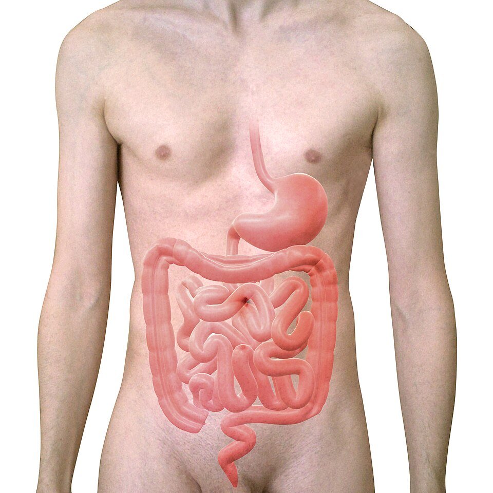
The Human intestine consists of two parts :
- Small intestine
- Large intestine
Small Intestine.
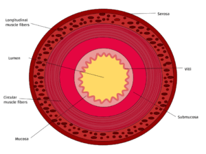
The small intestine is roughly 7 meters long. Like the stomach, its wall is also made of four layers.
- mucosa
- submucosa
- muscular layer
- peritoneal layer
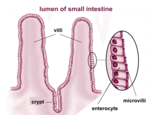
Almost all the chemical digestion takes place within the small intestine. Broken down food is now called nutrients, is also absorbed within the small intestine. In order to increase the surface area needed for absorption, the lining of intestinal walls has been equipped with finger-like projections called villi. The purpose of these villi is just to provide more and more surface area, needed for absorption. The villi are further covered with hair-like processes called microvilli, which further increase surface area. The surface of the small intestine has no natural protection against the aci,d, and this protection is provided by pancreatic juices. Chyme has now entered the small finger-like projections called villi. The purpose of these villi is just to provide more and more surface area, needed for absorption. The villi are further covered with hair-like processes called microvilli, which further
increase surface area. The surface of the small intestine has no natural protection against the acid, and this protection is provided by pancreatic juices. Chyme has now entered the small intestine. Most of the food is absorbed through villi and microvilli, whereas
absorption of fat is carried out through lacteals, present at the core of each villus. Absorb Fats and lipids take the form of a white milky fluid, giving them the name lacteal (milk). Lacteal fluid is named chyle.
The Small intestine is divided into three parts:
Dodenum.
It is the initial part of the small intestine. It is almost 25 cm long, a C-shaped part of the small intestine. The ampulla of Vater is about 4 inches from the pyloric opening. , formed by joining the hepatic and pancreatic ducts, coming out of the liver and pancreas, and joining together before
opening at this point. The duct formed by the joining of both these ducts is now known as the Hepatopancreatic duct. This part of the duodenum has no protection against the acid coming from the stomach. This acid is neutralized by pancreatic juices.
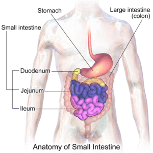
Jejunum.
It is the second part of the small intestine, they located between the duodenum and the ileum and is about 8 feet long. Most of the absorption occurs in this part of the small intestine. Villi and microvilli are present in maximum numbers throughout the length of the jejunum. The jejunum is the main site of nutrient absorption, and food is broken down into molecules.
- Carbohydrates are broken down into monosaccharides like glucose.
- Protein is broken down into many different amino acids.
- Fats absorb fatty acids and monoglycerides.
- Other minerals and salts also absorb, like water-soluble vitamins, fat-soluble vitamins, iron, calcium, etc..
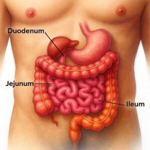
ILeum.
The ileum is the last portion of the small intestine and is about 12 feet long. Before reaching
the ileum, most of the absorption has already been completed. That is why the ileum has no or fewer
villi. The ileum is used to compact the leftover food. Food from the ileum is passed into
the large intestine through the caecum. The last part of the ileum doesn’t have the villi. After going through chemical digestion, nutrients from the food are absorbed through blood vessels present in the small intestinal walls by villi and microvilli and pass into the
portal circulation. Portal circulation transfers the nutrients to the liver, where these
nutrients are filtered and processed before being added to the bloodstream.
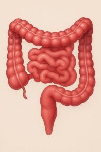
Large Intestine.
The final part of the digestive system is the large intestine. Its main function is the absorb water, salt, and electrolytes. The large intestine consists of three parts:
- Cecum.
-
Original AI-generated illustration by GetScienceMe – © 2025 Colon
- Rectum
The large intestine is anatomically divided into
- Ascending colon
- Transverse colon
- Descending colon
- Sigmoid colon
- Rectum
- Anus
Cecum.
It begins as the large intestine in the shape of a comma and connects the ileum at the ileocoecal junction. It is separated by a valve that does not permit the fecal matter to push upward and back into the ileum.
Colon.
Ascending Colon.
About 15 cm in length.Starts from the ecum in the right iliac fossa up to the lower end of the
Liver at the right colic flexure
Trensvers Colon.
It is about 50 cm in length. Starts from the right colic flexure to the left colic flexure, near
spleen.
Descending Colon.
About 12 cm long, starting from the left colic flexure to the sigmoid colon in the pelvis.
Sigmoid Colon.
The sigmoid colon is the S-shaped last part of the large intestine and is situated closest to the
rectum. It is about 35- 40 cm in size. The basic functions of the colon are to compact the waste
matter through absorbing leftover water and also to store and compact the fecal matter, and
make it ready to be evacuated through the rectum. The functions of the large intestine mainly include
the absorption of water. To some extent, bacterial digestion by normal flora ferments
the leftover carbohydrates. Vitamin B and Vitamin K are also synthesized here.
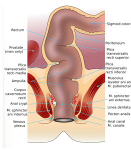
Rectum.
About 12 cm widened part of the large intestine is widened. It starts from the sigmoid colon to the anal canal. The function of the rectum is to store and expel feces.